Research

Our research on the structure and function of membrane proteins attacks one of the most important and challenging problems in contemporary biochemistry. Our most recent work has been focused on an outer membrane enzyme PagP from pathogenic Gram-negative bacteria. PagP transfers a palmitoyl group from a phospholipid molecule to the lipid A (endotoxin) component of lipopolysaccharide (LPS). This simple modification provides bacterial resistance to host antimicrobial peptides and attenuates the inflammatory response signaled through the human toll-like receptor 4 pathway. By understanding how the structural and dynamic properties of PagP relate to its biological function, we hope to design inhibitors that might be useful for the treatment of infections, and to synthesize novel endotoxin antagonists for the treatment of septic shock.

Figure 1

Figure 1

A Conformational perturbation hypothesis for PagP Gly88Cys.

Structural relationships between the non-degenerate exciton partners Tyr26 and Trp66 and Gly88 at the floor of the hydrocarbon ruler.

Mouse over the image to see the influence predicted for a Gly88Cys substitution.

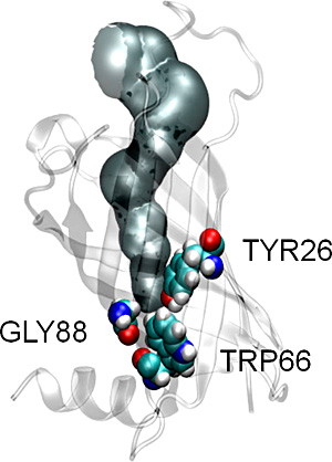
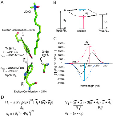
The structural basis of lipid acyl-chain selection by membrane-intrinsic enzymes is poorly understood because most integral membrane enzymes of lipid metabolism have proven refractory to structure determination; however, robust enzymes from the outer membranes of Gram-negative bacteria are now providing a first glimpse at the underlying mechanisms. The methylene unit resolution of the phospholipid:lipid A palmitoyltransferase PagP is determined by the hydrocarbon ruler, a 16-carbon saturated acyl-chain-binding pocket buried within the transmembrane β-barrel structure. Substitution of Gly88 lining the floor of the hydrocarbon ruler with Ala or Met makes the enzyme select specifically 15- or 12-carbon saturated acyl-chains, respectively, indicating that hydrocarbon ruler depth determines acyl-chain selection. However, the resolution of Gly88Cys PagP does not diminish linearly because it selects both 14- and 15-carbon saturated acyl-chains. We discovered that an exciton, emanating from a buried Tyr26-Trp66 phenol-indole interaction, is extinguished by a local structural perturbation arising from the proximal Gly88Cys PagP sulfhydryl group. Site-specific S-methylation of the single Cys afforded Gly88Cys-S-methyl PagP, which reasserted both the exciton and methylene unit resolution by specifically selecting 13-carbon saturated acyl-chains for transfer to lipid A. Unlike the other Gly88 substitutions, the Cys sulfhydryl group recedes from the hydrocarbon ruler floor and locally perturbs the subjacent Tyr26 and Trp66 aromatic rings. The resulting hydrocarbon ruler expansion thus occurs at the exciton’s expense and accommodates an extra methylene unit in the selected acyl-chain. The hydrocarbon ruler-exciton juxtaposition endows PagP with a molecular gauge to probe the structural basis of lipid acyl-chain selection in a membrane-intrinsic environment.

back to top
Figure 2

Figure 2

Theoretical analysis of a non-degenerate exciton interaction in PagP.

A) Structural relationships between the PagP hydrocarbon ruler, emphasizing the LDAO molecule and the pro-L hydrogen of Gly88, and the exciton interaction between Tyr26 and Trp66. The polarization axes for the interacting Tyr26 1Lai) and Trp66 1Bbj) electric transition dipole moment vectors are shown to emphasize their relationships with the interchromophore distance vector Rij. Key absorption parameters and the calculated exciton contribution to the wild-type PagP far-UV CD spectrum are also shown.
B) The π-π* transitions from ground states o to excited states a (for Tyr26 1La) or b (for Trp66 1Bb) are split by a non-degenerate exciton interaction into two new exciton states, which generate CD Cotton effects of equal magnitude, opposite sign, and separated by the splitting energy Δij. This splitting energy is given by (δij2 + 4Vij2)1/2 , where δij = (σi - σj) is the difference in the transition energies of the two chromophores, and Vij is the energy of interaction between the transition moments. Tyr26 and Trp66 each dominate the low (red) and high-energy (blue) exciton states, respectively.
C) The exciton couplet (solid curve) calculated from the Tyr26-Trp66 exciton interaction is the resultant of the two exciton-split CD Cotton effects (red and blue dotted curves), which are separated on the wavelength scale by Δij’, the wavelength equivalent of Δij.
D) The theoretical rotational strengths (Ro) of non-degenerate exciton split CD Cotton effects are proportional to Vij, Δij, and a scalar triple product involving the vector connecting the centers of the two transitions and the transition dipole moments. Vij can be approximated by the point-dipole approximation, but a more exact method is used in calculating the spectra shown here.

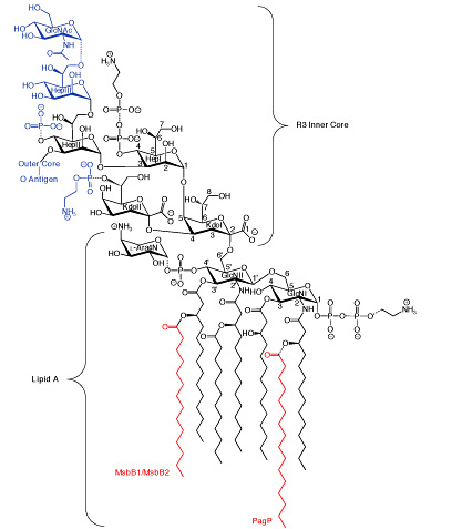
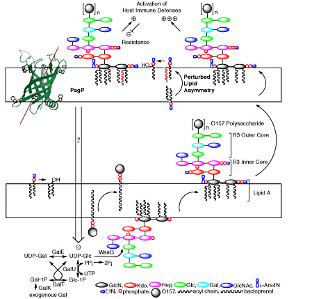
The enterobacterial outer membrane is an asymmetric lipid bilayer in which LPS exclusively lines the external leaflet while phospholipids line the inner leaflet. The asymmetric lipid organization provides a permeability barrier to hydrophobic antibiotics and detergents encountered in the natural and host environments. Although hydrophobic antibiotics can freely permeate through phospholipid bilayers, negative charges in LPS are bridged by Mg2+-ions to create tight lateral packing interactions that largely prevent permeation. Perturbations of outer membrane lipid asymmetry are thought to be associated with the migration of phospholipids into the external leaflet to create localized rafts of phospholipid bilayers, which render bacteria susceptible to hydrophobic antibiotics. Additionally, the LPS O-antigen can provide bacterial resistance to serum by preventing deposition of the complement cascade membrane attack complex.

back to top
Figure 3

Figure 3

Structure of lipid A and the inner R3 core oligosaccharide of E. coli O157:H7.

Lipid A is an acylated and phosphorylated disaccharide of GlcN linked by a β-1’,6-glycosidic bond. Three of the four primary R-3-hydroxymyristate chains can be modified with secondary acyloxyacyl groups. MsbB can incorporate a myristate chain and a palmitate chain can be partially incorporated by PagP (red). Partial modification of the lipid A phosphate groups with PEtN and L-Ara4N can also be observed. The inner core consists of two Kdo and three Hep sugars, which can be substituted with phosphate and PEtN groups. GlcNAc is a unique feature of the R3 inner core of E. coli O157:H7, but the remaining inner core structures and lipid A are virtually identical in E. coli K-12, which has a distinctly different outer core structure and no O-antigen. Structures shown in blue in the Hep domain are lost in E. coli O157:H7 as a consequence of an epistatic interaction between msbB and pagP.

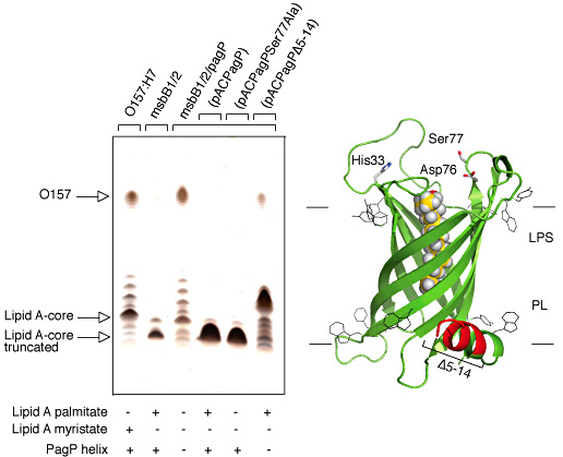
PagP structure and dynamics demonstrate that the palmitate recognition pocket, known as the hydrocarbon ruler, is only accessible from the outer membrane external leaflet and, thus, requires aberrant translocation of phospholipids into the external leaflet. Indeed, PagP remains dormant in the outer membrane until lipid redistribution associated with a breach in the permeability barrier directly triggers PagP activity. Lipid A palmitoylation is an appropriate adaptive response, because it helps to restore the permeability barrier. However, we have also shown that outer membrane activation of PagP can exert control on cytoplasmic enzymes of LPS core oligosaccharide structure that leads to a failure in O-antigen attachment. In this case, PagP does not require its catalytic machinery, but it depends instead on a periplasmic amphipathic α-helix.

Figure 4

Figure 4

Complementation of the truncated R3 core oligosaccharide of msbB-deficient E. coli O157:H7.

Silver stained 16% Tricine SDS-PAGE analysis of LPS isolated from wild-type E. coli O157:H7 strain 4303 and its lipid A acylation mutants deficient in either msbB or both msbB and pagP. Bacteria transformed with pACPagP, pACPagPSer77Ala, and pACPagPΔ5-14 are indicated above the figure. The presence (+) or absence (-) of either the PagP periplasmic amphipathic α-helix, or of lipid A acylated by palmitate or myristate, is indicated below each lane (left panel). Structural model of PagP derived from the crystal structure (PDB: 1THQ). The first seven N-terminal residues are disordered in the crystal structure, but the approximate position of the Δ5-14 deletion in the periplasmic amphipathic α-helix (red), and the cell surface catalytic residues, are indicated. The bound LDAO detergent molecule (yellow) identifies the location of the hydrocarbon ruler. The aromatic belts define the boundaries of the OM, where LPS occupies the external leaflet and phospholipids (PL) occupy the periplasmic leaflet (right panel).

back to top

The physical separation between the outer membrane and the cytoplasm dictates that PagP must be functioning as a sensory transducer, which is activated by a breach in the outer membrane permeability barrier. Our findings indicate that outer membrane activation of PagP not only triggers lipid A palmitoylation, but also separately triggers signal transduction across three cellular compartments. Discovering the downstream signaling components that respond to the activated-state of PagP will provide fertile ground for future research.

Figure 5

Figure 5

Model for PagP-mediated control of R3 core oligosaccharide structure.

The E. coli cell envelope includes both inner and outer membranes and the intervening periplasmic space. The primary enzymes of lipid A-core oligosaccharide and O-antigen biosynthesis require energy-rich metabolites and are confined to the cytoplasm or to the inner leaflet of the cytoplasmic membrane. Separate translocation events deliver the lipid A core and the bactoprenol-linked O-antigen subunit to the periplasmic face of the cytoplasmic membrane where the first extracytoplasmic reactions of LPS biosynthesis occur. O-antigen polymerization and ligation to the outer core is associated with partial modifications by the addition of PEtN, derived from PtdEtN to release diacylglycerol, and with L-Ara4N, derived from its bactoprenol-linked precursor (not shown). The entire LPS structure is then transported to the outer membrane (OM) and delivered to the external leaflet. The asymmetric OM lipid distribution, with LPS in the external leaflet and phospholipids in the inner leaflet, affords a permeability barrier to hydrophobic antibiotics and is compromised when phospholipids migrate aberrantly into the external leaflet. PagP is the only known OM enzyme of LPS biosynthesis in E. coli and it serves to transfer a palmitate chain from a phospholipid to lipid A with the production of a lysophospholipid by-product. Lipid A palmitoylation attenuates endotoxin signaling through the host TLR4 pathway and affords resistance to cationic antimicrobial peptides. PagP measures the phospholipid palmitate chain in its hydrocarbon ruler, which is only accessible from the OM external leaflet. A defect in cytoplasmic lipid A myristoylation triggers PagP activity in the OM of E. coli O157:H7. Activated PagP not only palmitoylates lipid A at the cell surface, to partially compensate for lipid A underacylation, but also separately initiates signal transduction through its periplasmic amphipathic α-helix. Signal transduction exerts negative control on the cytoplasmic UDP-Glc pool and leads to a truncation of the R3 core oligosaccharide.

back to top